
If the P wave morphology changes this may indicate a multifocal origin which is called wandering pacemaker. It is the first positive wave in the QRS complex.

Is A Narrow And Tall Qrs Complex An Ecg Marker For Sudden Death Heart Rhythm

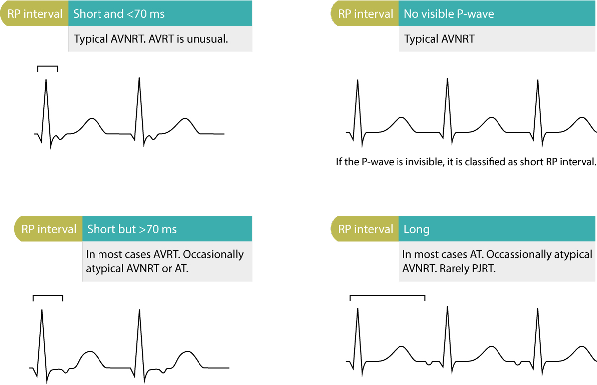
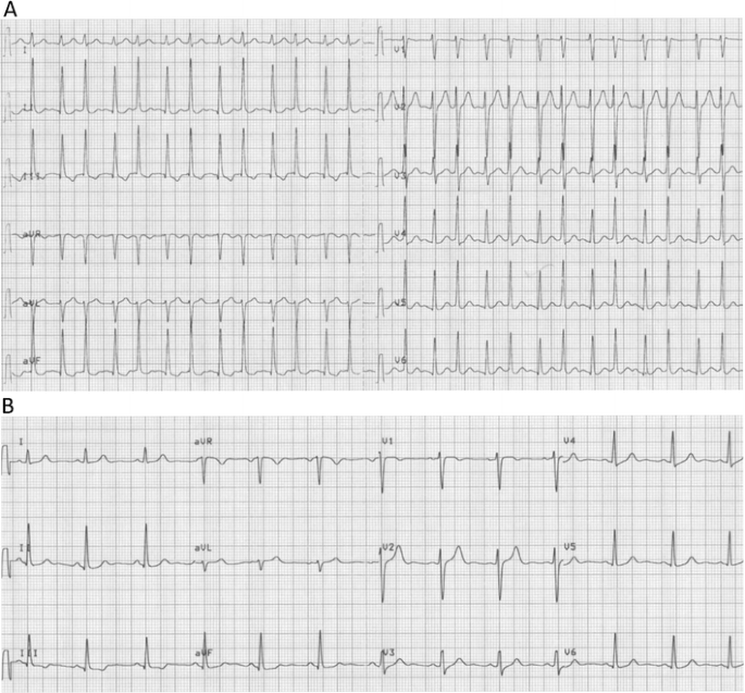
Ecg Shows Long Rp Narrow Complex Tachycardia With 1 1 Av Conduction P Download Scientific Diagram
Qrs Interval Litfl Ecg Library Basics
Narrow QRS rhythm suggests a junctional escape focus for the ventricles with block above the pacemaker focus usually in the AV node.
Narrow qrs complex. HeartRhythm the official Journal of the Heart Rhythm Society and the Cardiac Electrophysiology Society is a unique journal for fundamental discovery and clinical applicability. Regular wide complex tachycardia is most common and often represents VTach. What is the P-R interval.
Wide Complex Tachycardias WCTs are also known as Broad Complex or Wide QRS Complex Tachycardias. Most have a narrow QRS complex although occasionally electrical conduction abnormalities may produce a wide QRS complex that may mimic ventricular tachycardia VT. This initial distinction will guide the rest of the thinking needed to arrive at a final diagnosis.
A wide QRS complex especially left BBB is associated with more advanced myocardial injury worse left ventricular function and higher mortality than those in the case of a narrow QRS complex Figure 5. It can present different morphologies depending on the lead read QRS complex morphology. Betty Brown Created Date.
Narrow-complex capture beat complex 6 Several dissociated P waves are seen in the lead II rhythm strip associated with the 3rd 10th 14th 18th and 22nd QRS complexes This rhythm could easily be mistaken for SVT with bifascicular block RBBB LAFB however the presence of dissociated P waves and a narrow-complex capture beat indicates that this is fascicular VT arising from the. The QRS complex is the combination of three of the graphical deflections seen on a typical electrocardiogram ECG or EKGIt is usually the central and most visually obvious part of the tracing. At the end of this article is a quick quiz you can take to test your knowledge on measuring the QRS complex.
If the QRS duration is prolonged 012 seconds the arrhythmia is a wide complex tachycardia WCT. It consists of a collection of waves which represents the ventricular depolarisation. A narrow complex tachycardia is defined as a QRS complex.
The P-wave PR interval and PR segment. If the QRS duration is normal. They are narrow.
PR Interval WNL at first but lengthens progressively until P does not conduct to QRS Narrow. In adults the QRS complex normally lasts 80 to 100 ms. It ends at the point where the last wave of the complex transitions into the ST segment.
Due to bundle branch block hyperkalaemia or sodium-channel. Two studies showed that fQRS as well as wide QRS complex was associated with worse prognosis in patients with DCM 730. QRS complex starts where first wave of complex starts to move away from the baseline.
In this article I am going to explain how to easily measure the QRS complex. Is the R-R interval regular. Because the normal ventricular conduction system His-Purkinje is used the QRS complex is frequently narrow.
Get a full year access for only 26. Ventricular is fundamental since they are treated differently. Is the QRS wide or narrow.
QRS complex is larger than the P wave because ventricular depolarization involves a considerably larger muscle mass than atrial depolarization. Sinus atrial junctional or ventricular. If the first wave of the QRS complex is negative it is referred to as Q wave.
Its duration ranges from 006 s and 010 s. Is the R-R interval regular. The QRS width is useful in determining the origin of each QRS complex eg.
This group also includes antidromic AVRT and regular tachycardias with aberrancy. Rhythm Regularity Rate P Waves QRS Complex 2 AV Block Mobitz II Usually Regular May be Iregular. Aberrancy implies the patient has an EKG with baseline wide QRS from a bundle branch block BBB.
Is there a QRS complex for every P-wave. A junctional rhythm is normally slow less than 60 beats per minute. Close examination of QRS complex in various leads reveals that the terminal forces ie 2nd half of QRS.
012 second Vagal maneuvers if regular Adenosine if regular β-Blocker or calcium channel blocker Consider expert consultation Consider Adenosine only if regular and monomorphic Antiarrhythmic infusion Expert consultation If refractory consider Underlying cause. Broad complexes QRS 100 ms may be either ventricular in origin or due to aberrant conduction of supraventricular complexes eg. Is the QRS wide or narrow.
Master ECG interpretation from our nationally-known educators. HeartRhythm integrates the entire cardiac electrophysiology EP community from basic and clinical academic researchers private practitioners engineers allied professionals industry and trainees all. It is easiest to understand this nomenclature by considering these terms independently.
Normal q-waves reflect normal septal activation beginning on the LV septum. Are the QRS complexes grouped or not grouped. Narrow complexes QRS 100 ms are supraventricular in origin.
Atrial Fibrillation This rhythm is characterized by no waves before the QRS complex and a very irregular heart rate. Are the QRS complexes grouped or not grouped. What is the P-R interval.
Supraventricular tachycardia SVT is an extremely fast atrial rhythm with narrow QRS complexes when the impulse originates above the bundle branches above the ventricles. What is the QRS duration. The P-wave reflects atrial depolarization activation.
ECG interpretation traditionally starts with an assessment of the P-wave. The PR interval is the distance between the onset of the P-wave to the onset of the QRS complex. Betty Brown Created Date.
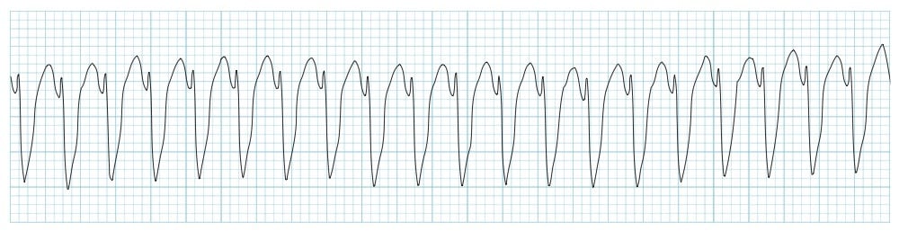
The QRS complex tends to be wide in ventricular tachycardia v-tach and very narrow in sinus tachycardia. The normal QRS complex during sinus rhythm is narrow. A compound this short illustrates the rapid activation of the ventricles which indicates.
Characterized by narrow QRS complexes preceded by P waves that do not fulfill one or more of the normal sinus rhythm NSR criteria mentioned earlier. The PR interval is assessed in order to determine whether impulse conduction from the atria to the ventricles is normal. If an old EKG is available the baseline wide QRS will be present.
In the clinical setting the distinction between narrow and wide complex tachycardia supraventricular vs. It corresponds to the depolarization of the right and left ventricles of the heart and contraction of the large ventricular muscles. For each QRS Complex Narrow 2 AV Block Mobitz I Wenckebach Regularly Irregular Usually 60 to 100 May be Bradycardic Positive.
Wide refers to a QRS complex duration width of greater than or equal to 012 seconds 120 msec corresponding to three small boxes on the ECG paper. Is there a QRS complex for every P-wave. They are often seen in leads I and aVL when the QRS axis is to the left of 60 and in leads II III aVF when the QRS axis is to the right of 60.
What is the QRS duration.
Aphrs2019thailand Com

Ecg Session Narrow And Wide Complex Tachycardia Youtube
Chicago Medicine Uic Edu
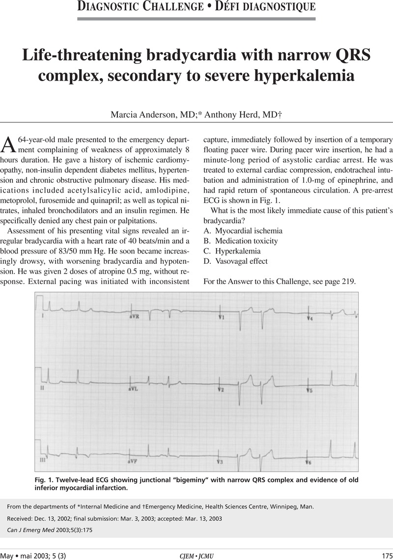
Life Threatening Bradycardia With Narrow Qrs Complex Secondary To Severe Hyperkalemia Canadian Journal Of Emergency Medicine Cambridge Core

A Ecg Showed An Irregular Narrow Qrs Complex Tachycardia Which Download Scientific Diagram
Diagnosis And Management Of Narrow And Wide Complex Tachycardia Ecg Echo
Overview Of Cardiac Arrhythmias Knowledge Amboss
A Supraventricular Tachycardia Showing Alternation Of The Qrs Interval Springerlink


